SARS-CoV2 and Vitamin D Interaction || Variant of SARS-CoV2 introduced
In my previous post, I explained the Pathophysiology and Pathogenesis of SARS-CoV-2, which is the Covid-19 virus. We are still on SARS-CoV-2, so on that note, we will be checking its relationship with Vitamin D. I checked through some articles that showed that people with low vitamin D have a 95% confidence interval [CI] for Covid 19, 1,2 low level of vitamin D associated with Covid-19 patient staying longer using ventilators.3 Also, other papers showed a higher Covid-19 PCRS being associated with lower vitamin D,4 also lower vitamin D level were reported to be associated with increased fibrinogen, CRP, D-Dimer levels in patients with Covid -19.5 Other experiments shows decrease clearance of SARS-CoV2 being associated with decreased vitamin D,6, 7, and lower vitamin D being associated with elderly, people with obesity,African American, and Hispanic races.8, 9, 10.

Vitamin D comes from exogenous sources such as the food we eat such as yoghurts, egg yolk, liver, mushrooms, and milk, and medications (supplements) known as Cholecalciferol. And this gets ingested through the Gastrointestinal tract. It is important to know that vitamin D has to bind with fat and cholesterol in other for it to be absorbed into the bloodstream because it is a fat soluble vitamin.11 Vitamin D, can also be gotten through the skin and it is absorbed through the cholesterol 7-dehydrocholesterol which is converted to Cholecalciferol under sunlight or UV-light. Vitamin D is a fat soluble hormone something like a steroid and it has to be bound to Vitamin-D Binding proteins which take them to their destination organs.12, 13. In the liver, 25-hydroxylase add an hydroxyl group to the 25th carbon of cholecalciferol, to become 25-hydroxyl cholecalciferol.13 It goes to the kidney where in the convoluted tubule 1α, hydroxylase puts an OH in the first carbon of the cholecalciferol, turning it into 1α,25-dihydroxycholecalciferol.14,15

The 1α,25-dihydroxycholecalciferol passes through the cell membrane where it binds with Vitamin D receptor Protein then to mRNA which then makes proteins Cathelicidins and Beta Defensins. In one word, vitamin D can act on the macrophages to increase Cathelicidins and Beta Defensins protein synthesis which is anti-microbial proteins. Beta Defensins puncture the envelope of the virus which is made up of a phospholipid bilayer. This mechanism works in Respiratory sensational viruses and influenza viruses. Cathelicidins cleave viral proteins on the envelope (proteolysis) that makes the virus, as well as stimulate macrophages to undergo phagocytosis of viral particles.16, 17, 18, 19,20,21
The Cytokine storm with SARS-CoV2 virus will cause multiple organ failure such as attacking the heart, causing myocardial infactions, heart failure, it will also act on the lungs leading o Acute Respitory Distress Syndrome (ARDS), the kidney where it would lead to Acute Kindey Injury, and the Liver increasing Arterial Sceloris, increasing thrombotic state and reactive oxygen species. With vitamin D, increased or normalized, there will increase in the destruction of the virus, there will be more macrophage activity, increasing the TH2 cell production of interleukin 10 which will decrease the cytokine storm, which will prevent multiple organ damage. 22, 23, 24.
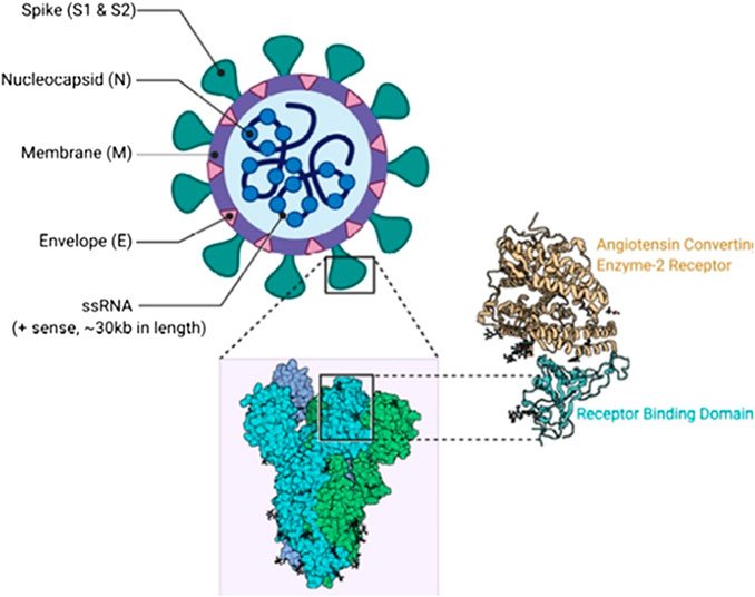
There are several variant of the SARS-CoV2 virus, so let's quickly look at them. The original SARS-CoV2 virus variant was the D614G 25, and it is important that we do not forget that the Spike protein is the protein of concern with the Covid-19 virus, although other proteins are important such as the Enveelope, and the Nucleo capsid. In the virus is the Single Stranded RNA genetic material. The original virus spike protein as I discussed in my previous post would bind to the ACE-2 protein of the alveoli which then causes a receptor mediated endocytocis. Inside the Cell, the RNA is released into the cytoplasm where it interact with both the free ribosomes and the ones with the Rough Endoplasmic reticulum. This allows for the creation of more Viral RNAs which will be used to create more new viruses. The gene responsible for creating the Spike protein is the concern with this variants as it mutation allows for another method of entry into the cell. The RNA polymerases expressed by the genes will be used to create more single stranded RNA. The RNAs and the viral protein are moved to the Golgi Aparatus where the virus starts to form in the vessicle. The Gulgi then fuses to the cell membrane where the viruses are released out of the cell, thereby destroying the cell and releasing new viruses to attach other cells.26, 27, 28
The viruses then mutate to become the B117 variant which is common in the UK and it is substitution between the N501Y, another variant is the B1351, the P1, and the Delta variants. In my next posts, I will explain these variants but it is important to know what causes these variants to be different.
Image Reference
Image 1 || Frontier || Contributed by Rohan Bir Singh, MD; Made with Biorender.com
Image 2 || brainandbodyfoundation.org || Vitamin D and Covid

Thanks for your contribution to the STEMsocial community. Feel free to join us on discord to get to know the rest of us!
Please consider delegating to the @stemsocial account (85% of the curation rewards are returned).
Thanks for including @stemsocial as a beneficiary, which gives you stronger support.